ANATOMIC PATHOLOGY SOFTWARE

LIS and Digital Pathology

Published by Esblada / Articles
The Pathology Information System as the heart of the digitisation project.
Continue reading

Implementing digital pathology without a laboratory information system (LIS) that is ready for it is like digitising the images and leaving the rest of the process on paper. The scanner captures the slide, but a diagnosis requires much more than just a high-resolution image.


What does the digitisation of anatomic pathology actually mean?

There is an important distinction that is often overlooked: digital pathology and the digitisation of the histopathology service are not the same thing. The former refers to the replacement of the microscope with high-resolution digital images for diagnostic purposes. The latter encompasses the entire process: from the moment the sample is received until the report reaches the requesting clinician.

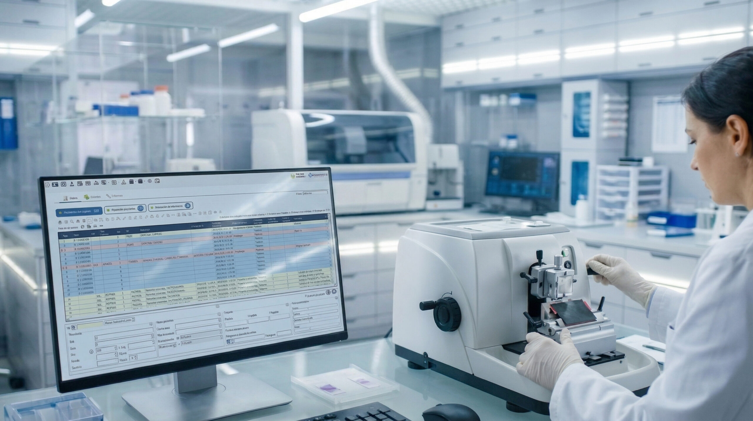
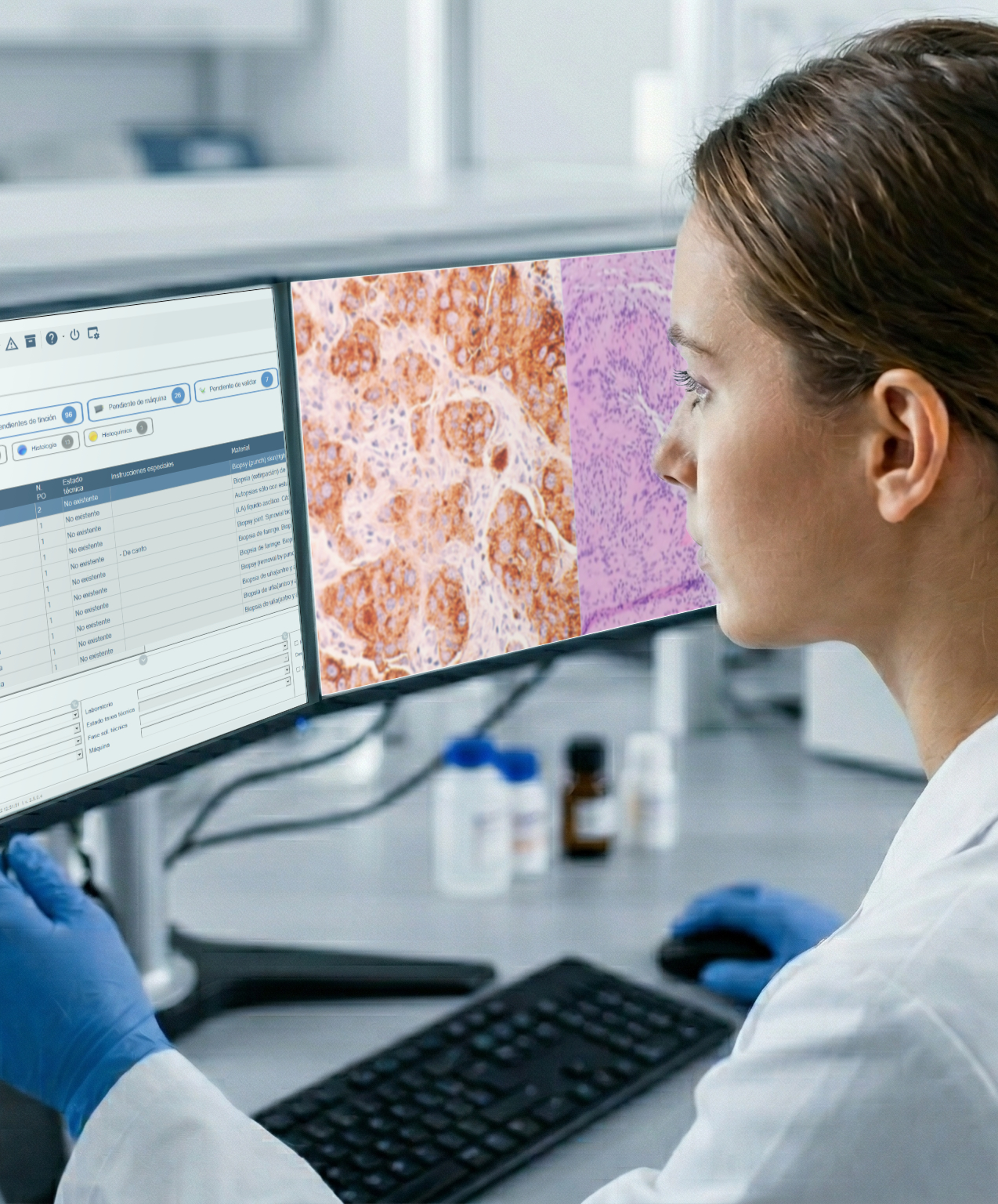
When a pathologist sits down at their screen to review a case, they need much more than just the scanned image of the slide. That image is the end result of a chain of processes that includes sample reception, paraffin embedding, sectioning, staining, scanning and technical validation. Any issue in that chain affects the diagnosis.

In order for the pathologist to make a confident diagnosis, they need access, within the same environment, to:

  • The patient’s clinical details and the context of the request
  • The type of sample, method of collection and conditions on receipt
  • The technical procedures carried out: number of sections, stains, special techniques
  • Any incidents recorded in the quality assurance process
  • The patient’s previous medical history and other relevant diagnostic test

All this information is managed by the LIS. Without an integrated information system offering full traceability, the pathologist has the image but not the context. And a histopathological diagnosis without context is an incomplete diagnosis.


The role of the LIS in a digital pathology project

The digital pathology projects currently being rolled out in regional communities and hospital networks have recognised this: the modernisation of the LIS and the introduction of the scanner go hand in hand. This is no coincidence. The fact is, they cannot function separately.
Integration with artificial intelligence for diagnosis

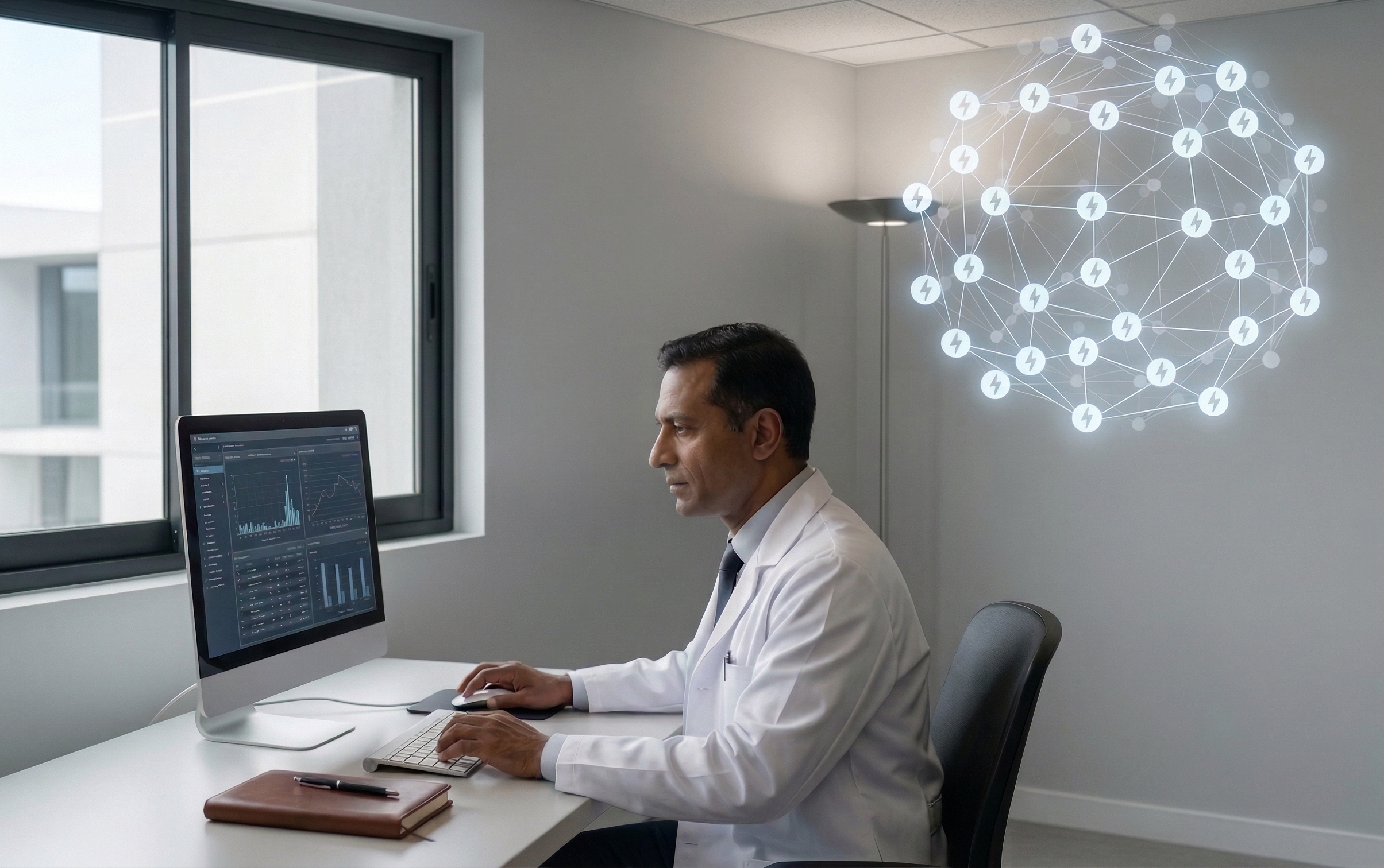
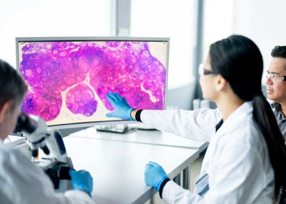
AI tools that analyse histological images require structured data to function effectively. The LIS provides this structure: sample type, previous diagnosis, stain used, protocol applied. Without this layer of information, the algorithms operate without clinical context, which limits their diagnostic utility.

Real-time multi-parameter searches

A repository of digitised cases with structured data enables cross-searches by diagnosis, morphology, technique, centre or time period. This has immediate value for day-to-day clinical practice, as well as for research and teaching.

Digitisation of the entire workflow

From the electronic request generated in the clinical system to the publication of the report in the patient’s record, the LIS manages every step of the process. This eliminates paper-based interruptions, reduces transcription errors and ensures that every action is logged with the user, date and time.

Sample traceability

The LIS maintains a complete record of all containers associated with the sample, all actions carried out on it and all equipment involved in the process. When the pathologist views the image, they know exactly what has happened to that tissue since it arrived at the laboratory.

Reports structured by protocol

The standardisation of diagnosis through synoptic protocols improves the quality of the report and facilitates the comparison of data between cases. It also forms the basis for any retrospective analysis and for integration with artificial intelligence tools.

Multisite work and case sharing

A LIS with a multisite architecture enables several hospitals to share cases, distribute the workload and maintain traceability between different locations. This is particularly useful in digital pathology networks where a single pathologist can review cases from multiple sites from a single workstation.

Which features of pathology software are key when digitising the department?

Not all information systems are suited to operate in a digital pathology environment. When evaluating a LIS for this context, it is advisable to check that it covers, as a minimum:

  • Native integration with the most common whole-slide image (WSI) scanners on the market
  • HL7/FHIR connectivity with the electronic health record system
  • Management of synoptic protocols (CAP, SEAP or others, depending on the centre)
  • Quality control module with real-time incident logging
  • Architecture designed for multi-site environments
  • Open API for integration with diagnostic AI platforms

Conclusion

Digital pathology changes where the diagnosis is made, but it does not eliminate any steps from the preceding process. Diagnostic value continues to depend on the quality of the specimen, the correct application of technical protocols and the availability of complete clinical information at the time of reading.

The LIS is the infrastructure that ensures all of this is available, organised and traceable. A digital pathology project that does not take the information system into account from the outset is not digitising the service: it is merely replacing the microscope with a screen.

Contact us

Sources and recommended reading

International publications — hospital experiences

  1. Temprana-Salvador J, López-García P, Castellví Vives J, et al. DigiPatICS: Digital Pathology Transformation of the Catalan Health Institute Network of 8 Hospitals — Planning, Implementation, and Preliminary Results. Diagnostics. 2022;12(4):852. https://doi.org/10.3390/diagnostics12040852
  2. Alchami FS, Iqbal Z, Björkhammer CN, et al. Whole Slide Imaging Integration with Lab Information Systems: A Study of the Requirements, Processes and Procedures Enabling a Reporting-Based Workflow. Pathology and Laboratory Medicine International. 2023;15:1–11. https://doaj.org/article/63905700db8f41688d93a289aef72a6a
  3. Fraggetta F, et al. Step-by-step digital pathology workflow implementation centred on the AP-LIS. Gravina Hospital, Caltagirone (Sicily, Italy). PubMed Central. https://pmc.ncbi.nlm.nih.gov/articles/8534326

International organisations

  1. College of American Pathologists (CAP). Cancer Reporting Tools / Electronic Cancer Protocols. Updated December 2024. https://www.cap.org/protocols-and-guidelines/cancer-reporting-tools/cancer-protocol-templates

share it on networks

Other Stories

digital
solutions
FOR ANATOMIC PATHOLOGY WORKFLOWS

From specimen reception to reporting, our solutions ensure end-to-end traceability, integration, and efficiency.
Esblada Medical
We are an agile, dynamic, and multidisciplinary team dedicated to customer satisfaction and service excellence.
Request catalogs

© 2026 / Esblada Medical / All rights reserved Security policy  /  Quality policy  /  Privacy  /  Legal  /  Cookies
Non-excepted cookies, own and third-party cookies, are used for analytical purposes and to show you personalized advertising based on a browsing profile created from your browsing habits (for example, pages visited). here.
Accept cookies Configure cookies Decline cookies
Basic cookie information
This website uses cookies and/or similar technologies that store and retrieve information when you browse. In general, these technologies can serve very different purposes, such as, for example, recognizing you as a user, obtaining information about your browsing habits or personalizing the way in which the content is displayed. The specific uses we make of these technologies are described below. By default, all cookies are disabled, except for technical ones, which are necessary for the website to function. If you wish to obtain more information or exercise your data protection rights, you can consult our Política de cookies".
Accept cookies Configure cookies
Technical cookies needed Always active
Technical cookies are strictly necessary for our website to work and for you to navigate through it. These types of cookies are those that, for example, allow us to identify you, give you access to certain restricted parts of the page if necessary, or remember different options or services already selected by you, such as your privacy preferences. Therefore, they are activated by default, your authorization is not necessary.rnThrough the configuration of your browser, you can block or alert the presence of this type of cookies, although such blocking will affect the proper functioning of the different functionalities of our website.
Cookies de análisis
Las cookies de análisis son las utilizadas para llevar a cabo el análisis anónimo del comportamiento de los usuarios de la web y que permiten medir la actividad del usuario y elaborar perfiles de navegación con el fin objetivo de mejorar los sitios web.
Confirm preferences
Contacta con nosotros
Controller: EMS Pathology Medical Solutions S.L. / Purpose: To respond to your requests for information. / Legal basis: Consent. / Recipients: Your personal data will not be disclosed to third parties unless required by law. / Data subject rights: You have the right to access, rectify and erase your data, as well as the right to data portability, restriction of processing and objection to processing, by contacting us at: info@esblada.com
Fill out the form to request a personalized demo
Controller: EMS Pathology Medical Solutions S.L. / Purpose: To respond to your requests for information. / Legal basis: Consent. / Recipients: Your personal data will not be disclosed to third parties unless required by law. / Data subject rights: You have the right to access, rectify and erase your data, as well as the right to data portability, restriction of processing and objection to processing, by contacting us at: info@esblada.com
Fill out the form to download the catalogs
Controller: EMS Pathology Medical Solutions S.L. / Purpose: To respond to your requests for information. / Legal basis: Consent. / Recipients: Your personal data will not be disclosed to third parties unless required by law. / Data subject rights: You have the right to access, rectify and erase your data, as well as the right to data portability, restriction of processing and objection to processing, by contacting us at: info@esblada.com
Aceptar